The presence of reflux is determined by the direction of flow because any significant flow toward the feet is suggestive of reflux. The GSV is usually small further down the calf.
 Ultrasound Examination Of The Patient With Primary Venous Insufficiency Sciencedirect
Although identification and quantification of venous reflux is the primary focus of the sonographic evaluation of venous insufficiency the presence and location of venous varicosities must also be documented.
Venous reflux ultrasound grading. Tortuous ureter with moderate dilatation. Reflux limited to the ureter. This combination of back pressure causes dilation and tortuosity of the veins ie varicosites.
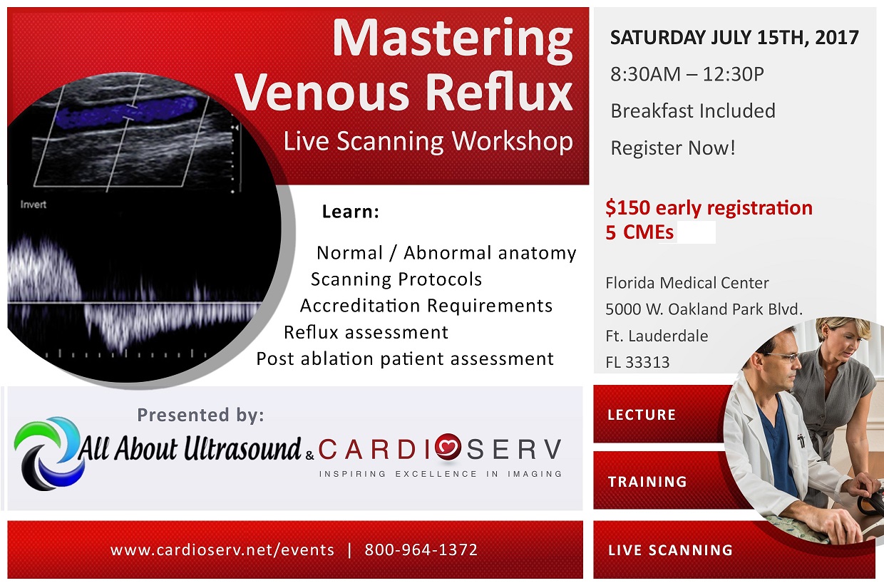
Venous Imaging for Reflux Using Duplex Ultrasonography Fig. Considered the primary imaging modality of choice. We will review the pathophysiology of venous reflux signs and symptoms ICAVL scanning protocols diagnostic criteria for ablation and much more at our upcoming Mastering Venous Reflux workshop.
Duplex ultrasound report of a patient with bilateral CVD who had venous claudication on the left LE and no symptoms on the right LE. Its designed to be very specific. Red flow positive signal going toward the transducer in the GSV is retrograde flow indicating reflux.
An incompetent perforating vein also allows blood to flow from the deep veins to the surface veins. Vesicoureteric reflux VUR grading divides vesicoureteric reflux VUR according to the height of reflux up the ureters and degree of dilatation of the ureters. We classified the patterns of reflux into four grades as follows.
The majority of varicosities are large with a diameter exceeding 4 mm palpable and tortuous. Reflux is commonly present when the vein. A long segment of reflux in a saphenous vein which correlates with symptoms and clinical findings of venous insufficiency is more important.
Patients with an unclear source of venous edema or leg pain consistent with venous hypertension should also undergo venous ultrasound. Typically the great saphenous vein and the small saphenous vein and their primary tributaries are assessed. Reflux was found in the POPV MGV PTV SSV two tributaries of SSV and a posterior calf perforator that measured 39mm.
Note large and refluxing perforators Remember GSV has numerous variations and may not be present and not all perforators will be visualized. PVU has a simplified approach regardless of experience level. All patients being considered for treatment of varicose veins or venous ulceration clinical etiologic anatomic pathophysiologic CEAP clinical stage C2C6 should undergo ultrasound of the venous system to determine the location and patterns of superficial and deep venous incompetence and of any deep venous obstruction.
This is generally at the lowest or most peripheral level of the primary incompetent segment. You need to see an experienced phlebologist for a clinical evaluation. Mild dilatation of ureter and pelvicalyceal system.
411 Reflux in the GSV in the lower thigh is demonstrated with color flow left panel and with Doppler waveform right panel. Incompetent valve perforator leading to venous reflux. Device may also be used to treat truncal venous reflux.
LOWER EXTREMITY SUPERFICIAL VENOUS REFLUX ULTRASOUND e-course - 60 CME Credits. GRADE 2B 2 We recommend open surgery is appropriate in veins not amenable to endovenous procedures but otherwise is not recommended because of increased pain convalescent time and morbidity. CEAP classification ranges from C0 no venous disease to C6 an open and active ulcer.
The size transition seen on ultrasound. Mastering Venous Reflux CME Workshop Ultrasound plays a vital role in identifying venous reflux. Ultrasound Venous Doppler ultrasound.
Groin tributaries and GSV were dilated and patent with continuous flow. In short it offers everything that the previous system failed to do. Today all healthcare professionals use the CEAP system when describing vein disease.
Knee measuring size and reflux just below knee mid calf and ankle. Venous insufficiency will get worse over time since we all live in a world where gravity creates pressure in veins in the leg. Lower-limb varicose veins are most often found along the trunk of the GSV.
Reflux up to the renal pelvis. In these cases the access will be at the level of the takeoff of this tributary. The superficial system is the most difficult venous system to ultrasound.
It then refluxes back down the leg through the malfunctioning valve. Focus on large and obviously refluxing veins. Duplex ultrasound is used first to identify the location for venous access.
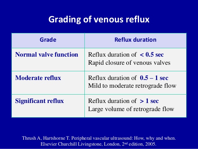
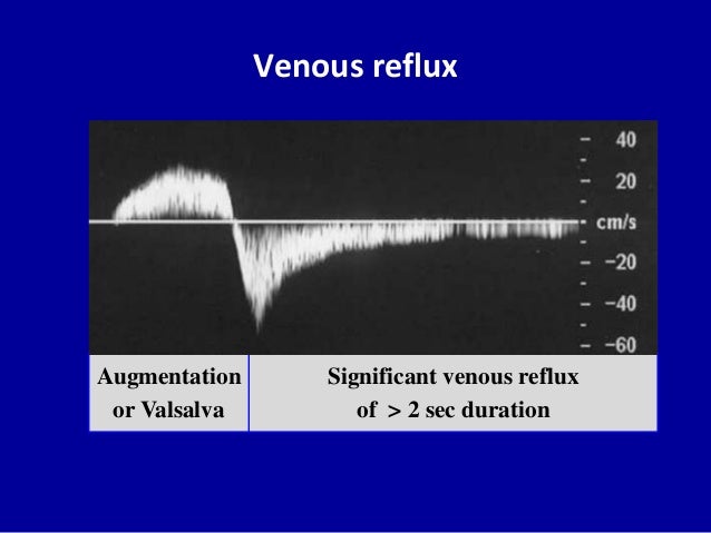
The duration of reflux is known as the reflux. 1 second of reflux is negative for valvular incompetence 1 second of reflux is positive for valvular incompetence This is our criteria. Typically the GSV refiuxes from the SFV down toward the calf where its incompetent fiow spills into a tributary that fills varicosities.
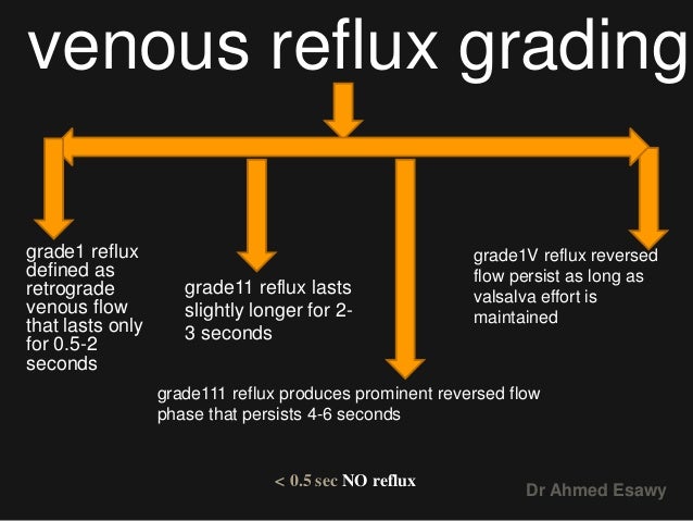
Find origin of Small saphenous veinSSV may be. Grade 1 retrograde Grade 2 augmented Grade 3 enhancing and Grade 4 stasis Table 1. There is something for everyone to learn.
 The 2020 Appropriate Use Criteria For Chronic Lower Extremity Venous Disease Of The American Venous Forum The Society For Vascular Surgery The American Vein And Lymphatic Society And The Society Of Interventional
 Lower Extremity Venous Duplex Ultrasound Chronic Venous Insufficiency Youtube
 Chronic Venous Insufficiency Circulation
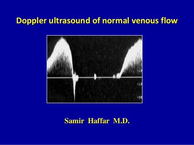 Doppler Ultrasound Of Normal Venous Flow
 4 Peripheral Venous Duplex Pt 4 Varices Dr Ahmed Esawy
 Prevalence Of Deep Venous Reflux As Primary Aetiology In Case Of Lower Limb Varicose Veins Abstract Id No Ppt Download
 Correlation Between Venous Reflux And Size With Venous Disability Scoring Download Table
 Superficial And Deep Venous Reflux Notes Reflux Is Measured While Download Scientific Diagram
Tips For Obtaining Accurate Venous Reflux
 Venous Insufficiency Exam Case Study Youtube
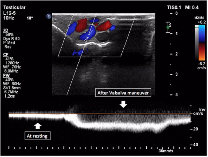 A Novel Method For Investigating The Role Of Reflux Pattern In Color Doppler Ultrasound For Grading Of Varicocele Scientific Reports

 Doppler Ultrasound Of Normal Venous Flow


No comments:
Post a Comment
Note: Only a member of this blog may post a comment.